Introduction
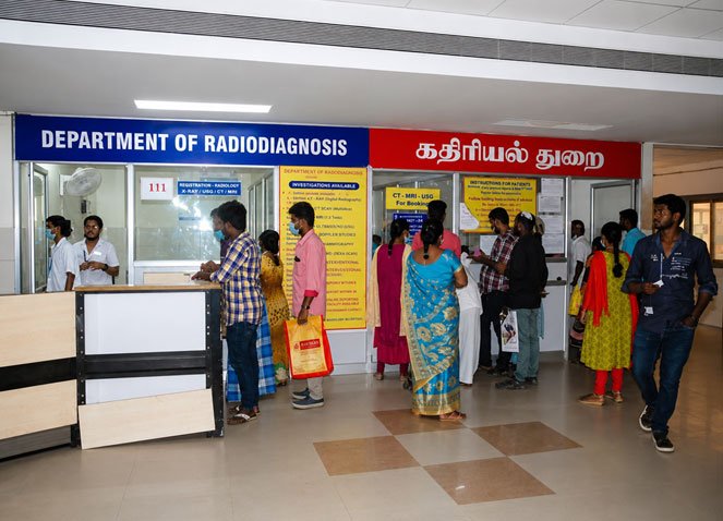
The Department of Radiodiagnosis at Government Mohan Kumaramangalam Medical College Hospital (GMKMCH), Salem was established in 1953 with the installation of the hospital's first X-ray unit, marking the beginning of comprehensive diagnostic imaging services for the people of Salem and the neighbouring districts.
The department has grown steadily over the decades by adopting advanced imaging technologies to meet the increasing healthcare needs of patients.
In 1989, Ultrasonography (USG) services were introduced. Today, the department operates 10 high-end ultrasound machines, all equipped with Colour Doppler facilities, performing an average of 180–200 ultrasound examinations daily. High-frequency Doppler ultrasound services were introduced in 2004, with 7–9 Doppler studies performed each day.
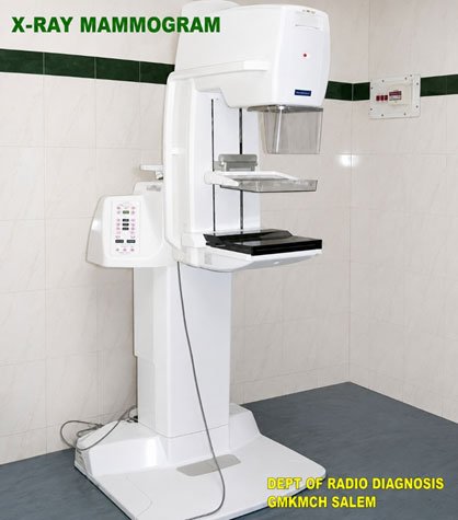
Mammography services were established in 2005 to support the early detection and diagnosis of breast diseases. The department currently performs 4–5 mammography examinations daily.
The department introduced its first Single-Slice CT Scanner in 1997, significantly enhancing diagnostic capabilities. Over the years, CT imaging services have expanded to include advanced multidetector CT technology. At present, the department performs 200–250 CT examinations daily, including 24×7 emergency CT services for trauma and critically ill patients.
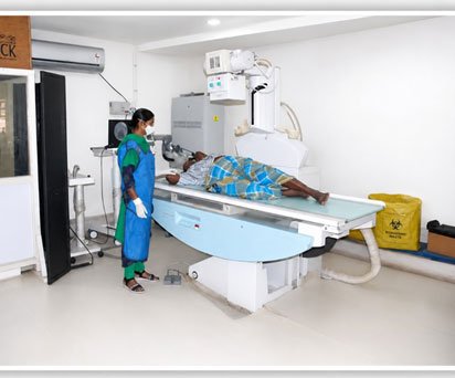
A major milestone was achieved in 2011, when the Department of Radiodiagnosis was relocated to the newly constructed PMSSY Super Speciality Block, enabling the introduction of several advanced diagnostic imaging services. An 800 mA X-ray unit with Image Intensifier Television (IITV) was commissioned to perform a wide range of fluoroscopic and contrast-enhanced diagnostic procedures.
Further technological advancements include:
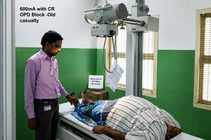
- 2015 – Installation of a 600 mA X-ray unit with Computed Radiography (CR) in the OPD Block.
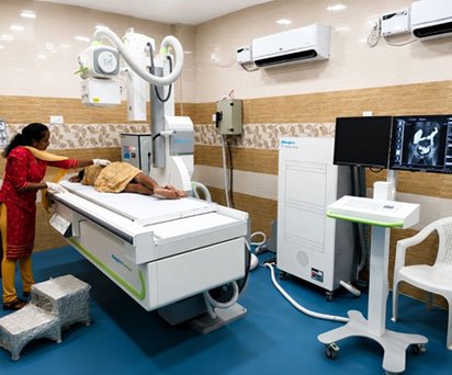
- 2015 – Installation of an 800 mA Digital Radiography (DR) system integrated with PACS in the PMSSY Block.
- 2019 – Commissioning of a new 16-Slice CT Scanner in the Medical Block. During the COVID-19 pandemic, this scanner played a vital role in diagnosing and triaging thousands of patients.
- 2021 – Replacement of the existing MRI system with a state-of-the-art 1.5 Tesla MRI scanner, equipped with advanced imaging applications for high-quality neurological, musculoskeletal, abdominal, vascular, and oncological imaging. The department currently performs 25–30 MRI examinations daily.
- 2021 – Installation of two advanced 3D/4D Ultrasound Systems with Elastography, enhancing diagnostic accuracy in obstetrics, abdominal imaging, and liver disease assessment.
- 2022 – Installation of a modern 800 mA Digital Fluoroscopy System with IITV in the Medical Block, strengthening the department's capabilities in contrast-enhanced and interventional radiological procedures.
Today, the Department of Radiodiagnosis is one of the leading diagnostic imaging centres in the region, offering comprehensive imaging services including Digital X-ray, Fluoroscopy, Mammography, Ultrasound & Colour Doppler, Computed Tomography (CT), Magnetic Resonance Imaging (MRI), PET-CT, Interventional Radiology, and Bone Mineral Density (DEXA) studies.
With advanced imaging technology, experienced faculty, skilled technical staff, and a commitment to quality patient care, the department provides round-the-clock diagnostic services to outpatients, inpatients, emergency and trauma cases, while also serving as a major referral centre for Salem and the neighbouring districts of Namakkal, Dharmapuri, Krishnagiri, Karur, Kallakurichi, and surrounding regions.
Faculty Details
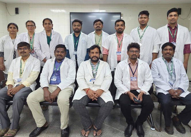
The Department of Radiodiagnosis is led by experienced faculty members dedicated to excellence in patient care, medical education, and research.
| S.No | Designation | Faculty Strength | Faculty Members |
|---|---|---|---|
| 1. | Professor and HOD | 1 | Dr. P. Kumar, MD (Radiodiagnosis) |
| 2. | Associate Professors | 2 | Dr. N. Sundareswaran, MD (Radiodiagnosis) Dr. M. Sankar, MD (Radiodiagnosis) |
| 3. | Assistant Professors | 7 | Dr. E. Vidhya, DMRD, DNB (Radiodiagnosis) Dr. P. Ramchandran, MD (Radiodiagnosis) Dr. M. Pradeepvel, MD (Radiodiagnosis) Dr. S. Nithiyan, MD (Radiodiagnosis) Dr. A. S. Kannan, MD (Radiodiagnosis) Dr. N. Ramkumar, MD (Radiodiagnosis) Dr. R. Sasikumar, MD (Radiodiagnosis) |
| 4. | Senior Residents | 4 | Dr. M. Shalini, MD (Radiodiagnosis) Dr. S. Ramakrishnan, MD (Radiodiagnosis) Dr. A. Dheepa, DMRD Dr. G. Jaysiya, DMRD |
Department Highlights
- Experienced and highly qualified faculty members
- Expertise in Diagnostic Radiology and Advanced Imaging
- Dedicated to Undergraduate and Postgraduate Medical Education
- Comprehensive Clinical and Academic Training
- Active participation in Research, Continuing Medical Education (CME), and Interdisciplinary Patient Care
- Round-the-clock diagnostic support for emergency and trauma services
Address for Communication
For enquiries, academic correspondence, and departmental communication, please contact:
The Registrar, Department of Radiodiagnosis
Government Mohan Kumaramangalam Medical College Hospital (GMKMCH),
Salem – 636001, Tamil Nadu, India.
Email: radiodiagnosissalemgh@gmail.com
Registrar: Dr. P. Ramachandran, MD (Radiodiagnosis)
Phone: +91 99441 58524
Emergency Radiology Services: Available 24 × 7
Contact Us
For academic enquiries, referral information, or diagnostic services, please contact the Department of Radiodiagnosis during office hours or visit the department at GMKMCH, Salem.
Post Graduate Students (MD Radiodiagnosis)
The Department of Radiodiagnosis, GMKMCH Salem, provides postgraduate training in Radiodiagnosis with structured academic teaching, clinical training, and exposure to advanced imaging modalities.
PG Batch Details
| Academic Year | Year of Training | Post Graduate Students |
|---|---|---|
| 2023 – 2026 Batch | Third Year PG | Dr. S. Sandhanaa Dr. B. Mahalakshmi Dr. K. Nithika |
| 2024 – 2027 Batch | Second Year PG | Dr. R. Ragavi Dr. Cherian S. Valliyammel Dr. P. Badhri Narayana |
| 2026 – 2028 Batch | First Year PG | Dr. Sangameshwar Barole Dr. E. Swetha Dr. R. Vimal Kumar |
Postgraduate Training Highlights
- Comprehensive training in Diagnostic Radiology
- Exposure to X-ray, Ultrasound, Doppler, CT, MRI, PET-CT, Mammography, and Interventional Radiology
- Regular academic sessions, seminars, case discussions, and journal reviews
- Clinical training under experienced faculty members
- Hands-on experience in emergency and advanced imaging services
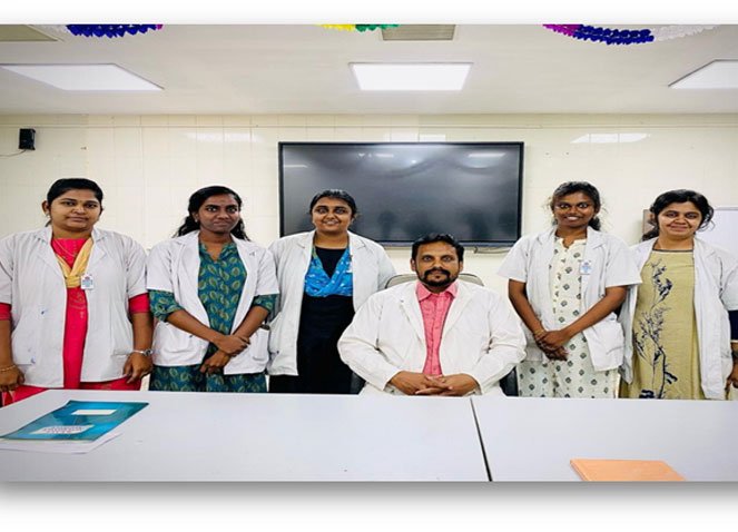
Technical Staff
The Department of Radiodiagnosis is supported by a dedicated team of technical and support staff who ensure smooth functioning of all diagnostic imaging services including X-ray, CT, MRI, Fluoroscopy, Ultrasound, Mammography, PET-CT, and emergency imaging services.
Technical Staff Distribution
| Category | Staff Strength |
|---|---|
| Radiographers | 14 |
| Dark Room Assistants (DRA) | 16 |
| Data Entry Operators | 6 |
| Supporting Staff | 1 |
| Total Technical & Support Staff | 37 |
Role of Technical Staff
Radiographers
Perform patient positioning and image acquisition for:
- Digital X-ray
- CT Scan
- MRI
- Fluoroscopy
- Mammography
- Emergency radiological procedures
Provide 24×7 imaging support for emergency and inpatient services.
Dark Room Assistants (DRA)
- Assist in imaging workflow and patient preparation.
- Support radiographers and radiologists in routine and emergency diagnostic services.
- Maintain smooth patient movement and departmental operations.
Data Entry Operators
- Support patient registration and digital data management.
- Assist in report processing and PACS-based workflow.
- Maintain accurate imaging records and documentation.
Supporting Staff
- Provide patient assistance and departmental support services.
Highlights
- Dedicated technical team supporting advanced imaging facilities.
- 24×7 emergency radiology service support.
- Trained personnel for CT, MRI, X-ray, Ultrasound, Fluoroscopy, and Interventional Radiology services.
- Efficient coordination between radiologists, clinicians, and technical staff for quality patient care.
Services Offered
The Department of Radiodiagnosis provides comprehensive diagnostic imaging services with advanced imaging equipment and experienced specialists. The department caters to outpatient, inpatient, emergency, trauma, and super-specialty services, offering high-quality diagnostic support 24×7.
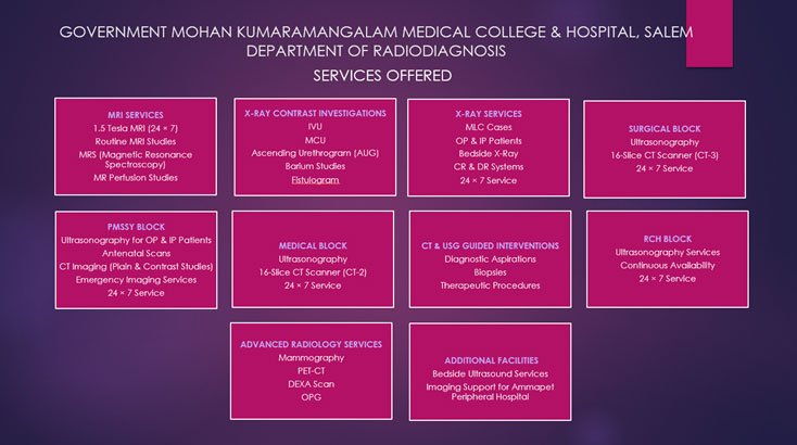
1. X-Ray
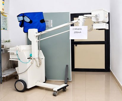
The department is equipped with 4 fixed X-ray units and 6 mobile X-ray units, providing routine and emergency radiographic services.
- Average workload: 400–450 X-ray examinations per day
- Digital and Computed Radiography (CR) facilities available
- Mobile X-ray services for ICU, emergency, trauma, and bedridden patients
| Machine | Strength | Fixed | Mobile |
|---|---|---|---|
| 800mA with IITV Recording System | 800mA | 2 | - |
| 800mA Digital Radiography - CEG - 1 | 800mA | 1 | - |
| 500mA with CR Systems | 500mA | 2 | - |
| 300mA with CR System | 300mA | 2 | - |
| 100mA Mobile Unit | 100mA | - | 6 |
| X-ray Mammo Gram Unit | - | 1 | - |
| 1000mA DR in Medical Block | 1000mA | 1 | - |
2. Fluoroscopy
The department has modern fluoroscopy units for diagnostic and contrast imaging procedures.
- Average workload: 5–8 procedures per day
- 800 mA IITV Fluoroscopy Unit (PMSSY Block)
- 800 mA Digital Radiography (DR) Fluoroscopy Unit (Medical Block)
Procedures Performed
- Barium Swallow
- Barium Meal
- Barium Follow Through
- Barium Enema
- Intravenous Urography (IVU)
- Micturating Cystourethrogram (MCU)
- Loopogram
- T-Tube Cholangiography
- Other Contrast Studies
3. Mammography
The department provides breast imaging services for screening and diagnosis.
- Average workload: 5–6 mammography examinations per day
- Digital Mammography for early detection of breast diseases
- Diagnostic and screening mammograms
5. Ultrasound
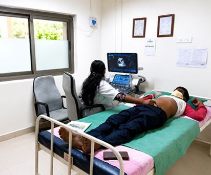
There are around 9 to 10 well equipped ultrasound Doppler machines with high frequency probes are available in our department. Now an average of 150 to 200 cases of ultrasound are done routinely.
We render separate ultrasound services for Out patients, Inpatients, Antenatal patients, doppler studies, super speciality IN & OUT patients, emergency - trauma care services and USG guided biopsy - procedures.
Two advanced MINDRAY DC80 machines installed in 2021 with 4D probe and recent elastography techniques has supported the patients in diagnosing better.
6. Computed Tomography (CT)
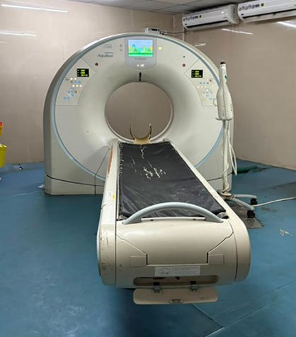
The department provides round-the-clock CT imaging services using advanced multidetector CT scanners.
- 24×7 Emergency CT Services
- Average workload: Approximately 200 CT examinations per day
- 15–20 Contrast CT studies per day
- CT-guided biopsy and interventional procedures
- During the COVID-19 pandemic, the dedicated CT unit performed over 12,000 CT examinations, significantly supporting patient diagnosis and management.
CT Services
- Routine CT
- Emergency CT
- Trauma CT
- CT Angiography
- Contrast CT
- CT-Guided Biopsy
- CT-Guided Drainage Procedures
| S.No | Machine | Company | Location |
|---|---|---|---|
| 1. | CT 16 - Slice | Aquilian Lightening | Speciality Block |
| 2. | CT 16 - Slice | Aquilian Lightening | Medical Block |
| 3. | CT 16 - Slice | Aquilian Lightening | Surgical Block |
7. Magnetic Resonance Imaging (MRI)
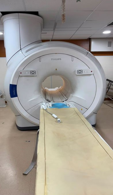
The department is equipped with a state-of-the-art 1.5 Tesla MRI scanner with advanced imaging applications.
Average workload: 30–40 MRI examinations per day
MRI Services
- Brain MRI
- Spine MRI
- Whole Body MRI
- Contrast MRI
- MR Angiography (MRA)
- MR Urography
- MR Spectroscopy
- MR Perfusion Imaging
8. Interventional Radiology
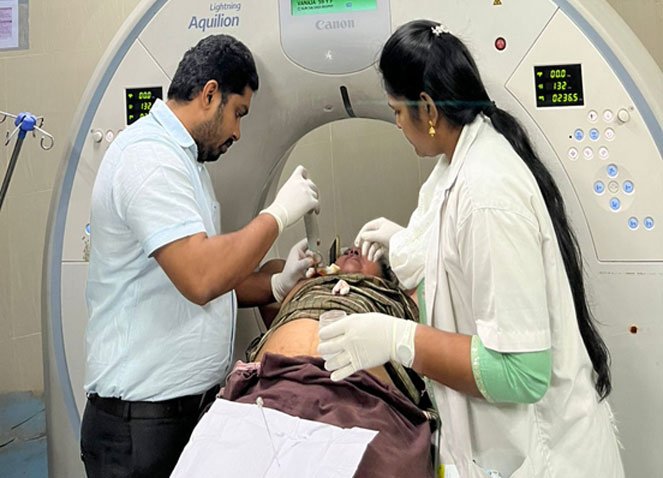
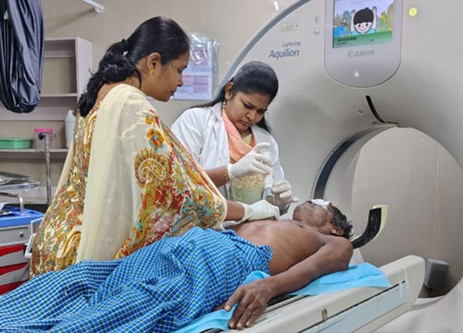
The department has a dedicated interventional suite for image-guided diagnostic and therapeutic procedures.
Average workload: More than 10 procedures per day
Procedures
- Ultrasound-Guided Biopsy
- CT-Guided Biopsy
- FNAC
- Ultrasound-Guided Aspiration
- Abscess Drainage
- Pigtail Catheter Insertion
- Liver Abscess Drainage
- Nephrostomy Procedures
- Thyroid FNAC
- Breast FNAC
The department is equipped with biopsy guns and coaxial biopsy needles for minimally invasive procedures.
Safety Standards
- All X-ray and CT equipment are licensed by the Atomic Energy Regulatory Board (AERB).
- All ultrasound machines are registered under the PCPNDT Act.
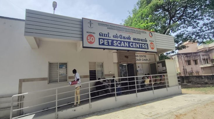
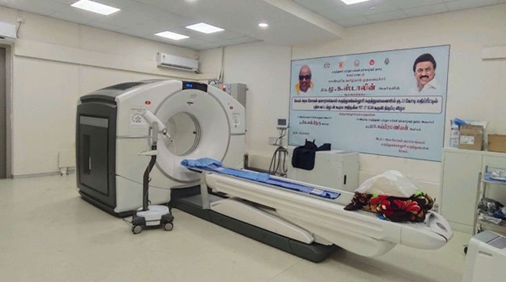
9. PET-CT
The Department of Radiodiagnosis is equipped with a state-of-the-art Positron Emission Tomography–Computed Tomography (PET-CT) scanner, providing advanced molecular and anatomical imaging for accurate diagnosis, staging, treatment planning, and follow-up.
PET-CT combines metabolic and structural imaging, enabling early disease detection and improving clinical decision-making.
Services
- Whole Body FDG PET-CT
- Oncology PET-CT
- Cancer Staging and Restaging
- Treatment Response Assessment
- Neurological PET Imaging
- Cardiac PET Imaging (where indicated)
- Infection and Inflammatory Disease Imaging
- Detection of Recurrent and Metastatic Disease
10. Bone Mineral Density (DEXA)
A Bone Densitometer (DEXA Scan) is available at the Master Health Check-up Unit.
Services
- Bone Mineral Density Assessment
- Osteoporosis Screening
- Fracture Risk Evaluation
- Follow-up of Osteoporosis Treatment
Highlights
- 24×7 Emergency Radiology Services
- PET-CT, MRI, CT, Ultrasound, Mammography & DEXA
- Three 16-Slice CT Scanners
- Advanced 1.5 Tesla MRI
- 9–10 High-End Ultrasound & Colour Doppler Systems
- Digital X-Ray (CR & DR)
- Image-Guided Interventional Procedures
- Experienced Radiologists & Technical Staff
- Serving Salem and Neighbouring Districts
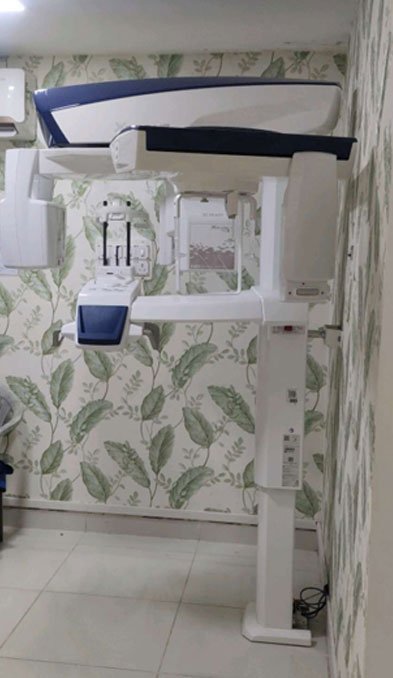
11. Orthopantomogram (OPG)
The Orthopantomogram (OPG) service was introduced in 2026. OPG is a digital panoramic X-ray that provides a comprehensive view of the teeth, jaws, and surrounding structures in a single image, aiding in the diagnosis and treatment planning of dental and maxillofacial conditions.
12. Contrast-Enhanced Mammography (CEM)
The Contrast-Enhanced Mammography (CEM) service was introduced in 2026. CEM is an advanced breast imaging technique that combines digital mammography with intravenous contrast to improve the detection and evaluation of breast abnormalities, particularly in women with dense breast tissue.
Daily Average Census of Various Sections
The Department of Radiodiagnosis provides comprehensive diagnostic imaging services to outpatients, inpatients, emergency cases, and referrals from various specialties. The department handles a high volume of diagnostic studies every day with advanced imaging facilities and dedicated technical support.
Daily Average Workload
| S.No | Section / Service | Average Daily Census |
|---|---|---|
| 1. | X-Ray Examinations | 400-500 |
| 2. | Special Procedures (IVP, Contrast Studies, etc.) | 5-10 |
| 3. | Ultrasonography & Doppler Studies | 200-250 |
| 4. | Computed Tomography (CT) Scans | 200-250 |
| 5. | Magnetic Resonance Imaging (MRI) Scans | 30-40 |
| 6. | Mammography | 4-5 |
| 7. | Interventional Radiology Procedures | 10-15 |
Department Highlights
- Total average daily imaging workload: Approximately 800-900 procedures/day
- 24×7 emergency imaging services available for trauma, critical care, and emergency patients.
- Advanced diagnostic facilities including:
- Digital X-Ray
- Ultrasound & Colour Doppler
- CT Scan
- MRI
- Mammography
- PET-CT
- Image-guided Interventional Procedures
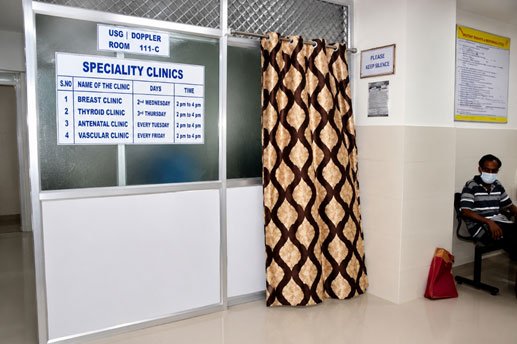
Special Clinics Conducted by the Department
The Department of Radiodiagnosis provides dedicated imaging support for various specialty clinics to facilitate early diagnosis, treatment planning, and follow-up care. These specialized clinics function in coordination with clinical departments and provide focused diagnostic services for patients.
Special Clinic Schedule
| Name of the Clinic | Day / Frequency | Timings | Clinic In-charge |
|---|---|---|---|
| Vascular Clinic | Every Friday | 8:00 AM – 11:00 AM | Dr. S. Ramakrishnan |
| Antenatal Clinic | Every Tuesday | 8:00 AM – 12:00 PM | Dr. A. Sharmila |
| Breast Clinic | 2nd Wednesday of Every Month | 8:00 AM – 11:00 AM | Dr. G. Jaysiya |
| Thyroid Clinic | 3rd Thursday of Every Month | 9:00 AM – 12:00 PM | Dr. A.S. Kannan |
Radiation Protective Measures
The Department of Radiodiagnosis follows strict radiation safety protocols to ensure the protection of patients, healthcare workers, and the public during diagnostic imaging procedures.
A full-time Radiation Safety Officer (RSO) cum Medical Physicist is posted in the department to oversee radiation protection practices, equipment safety, quality assurance, and compliance with regulatory requirements.
Radiation Safety Measures Implemented
- Continuous monitoring of radiation safety practices for patients, staff, and equipment.
- All X-ray and CT units are installed, tested, and certified according to the guidelines of the Atomic Energy Regulatory Board (AERB).
- Regular quality assurance and performance checks are carried out for radiological equipment.
- Thermoluminescent Dosimeter (TLD) badges are provided to all radiation workers for monitoring occupational radiation exposure.
- Radiation protection measures are implemented as per Bhabha Atomic Research Centre (BARC) specifications.
Radiation Protective Equipment Available
The department is equipped with appropriate radiation protection accessories, including:
- Lead protective screens
- Lead aprons
- Thyroid shields
- Gonadal protective guards
- Lead goggles
- Lead gloves
Commitment to Radiation Safety
The department follows the principle of ALARA (As Low As Reasonably Achievable) to minimize radiation exposure while maintaining high-quality diagnostic imaging.
Through proper monitoring, protective equipment, trained personnel, and adherence to AERB guidelines, the department ensures safe and effective radiological services for all patients and staff.
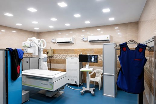
Department Library and Museum
The Department of Radiodiagnosis has a well-established departmental library and museum to support academic activities, postgraduate training, research, and continuous medical education.
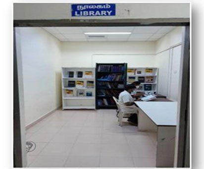
Department Library
The departmental library is well-furnished and equipped with internet facilities, providing access to updated academic resources for faculty members, postgraduate students, and trainees.
Library Facilities
| Category | Details |
|---|---|
| Total Number of Books | 164 |
| Latest Edition Books Purchased (Last 3 Years) | 87 |
| Internet Facility | Available |
| Academic Resources | Textbooks, Reference Books, Journals |
Journals Available
| Category | Number of Journals |
|---|---|
| Indian Journals | 08 |
| International Journals | 04 |
| Total | 12 |
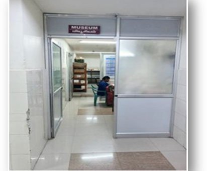
Department Museum
The department has a spacious and well-maintained museum facility supporting undergraduate and postgraduate teaching.
Museum Resources
- More than 300 specimens available for academic demonstration.
- More than 20 charts and diagrams illustrating radiological anatomy, pathology, and imaging concepts.
- Provides valuable learning resources for students and trainees.
- Supports practical teaching and case-based discussions.
Academic Highlights
- Dedicated learning environment for UG and PG students.
- Regular use of library resources for seminars, case presentations, and research activities.
- Museum facilities supporting systematic understanding of radiological findings and disease patterns.
Postgraduate (PG) Course
The Department of Radiodiagnosis offers postgraduate training in MD Radiodiagnosis with the objective of developing competent radiologists through comprehensive clinical training, academic teaching, research activities, and exposure to advanced imaging modalities.
The MD Radiodiagnosis degree course with an annual intake of 3 seats was started in the academic year 2022–2023.
The postgraduate training programme includes structured teaching, case discussions, seminars, journal reviews, practical demonstrations, and hands-on experience in various imaging modalities including X-ray, Ultrasound, Doppler, CT, MRI, PET-CT, Mammography, and Interventional Radiology.
PG Academic Activities
Regular academic activities are conducted for postgraduate students to enhance theoretical knowledge, clinical skills, and diagnostic expertise.
| S.No | Academic Activity | Schedule |
|---|---|---|
| 1. | Theory Classes | Daily morning 8:00 AM – 8:20 AM |
| 2. | Clinical Seminars | Every Wednesday |
| 3. | Journal Clubs | Every Saturday |
| 4. | Tutorials | Afternoon 12:00 PM – 1:00 PM |
| 5. | Film Reading Sessions | Daily afternoon 12:00 PM – 1:00 PM |
| 6. | Guest Lectures | Three times annually |
Academic Training Programmes
The Department of Radiodiagnosis has been actively involved in training medical professionals and paramedical personnel through various academic programmes.
Paramedical Courses
- CRA (Certificate in Radiological Assistant) Course - Started in 2008
- DRA (Diploma in Radiological Assistant) Course - Started in 2008
- DRDT (Diploma in Radiodiagnostic Technology) Course - Started in 2011
Ultrasound Training Programmes
The department provides structured ultrasound training throughout the year for:
- Medical Officers from Primary Health Centres (PHCs)
- Assistant Surgeons from Government Hospitals
- Doctors from ESI Hospitals
The training focuses on basic ultrasound techniques, image interpretation, patient preparation, and safe ultrasound practices.
Undergraduate & Postgraduate Teaching
The department conducts basic and advanced radiology teaching sessions for:
- Pre-final year MBBS students
- Final year MBBS students
- Postgraduate students
- Postgraduate trainees from other clinical specialties:
- General Surgery
- General Medicine
- Orthopaedics
- Obstetrics & Gynaecology
Training includes exposure to:
- Basic radiological anatomy
- Common imaging findings
- Appropriate selection of imaging modalities
- Clinical correlation of radiological findings
PCPNDT Ultrasound Training Programme
A six-month PCPNDT-compliant Ultrasound Training Programme was conducted for service and non-service medical officers from: December 2015 to May 2016
The programme provided training in:
- Basic ultrasound principles
- Obstetric ultrasound
- Abdominal ultrasound
- Ultrasound reporting practices
- PCPNDT Act guidelines and compliance
Department Academic Highlights
- Structured PG teaching programme
- Regular seminars and case discussions
- Journal club activities
- Film interpretation sessions
- Training in advanced imaging modalities
- Continuous medical education activities
- Skill development programmes for doctors and technical personnel
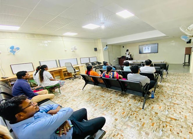
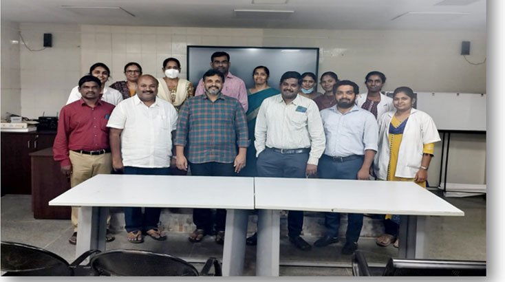
One-month USG training given to service medical officers from December 2023.
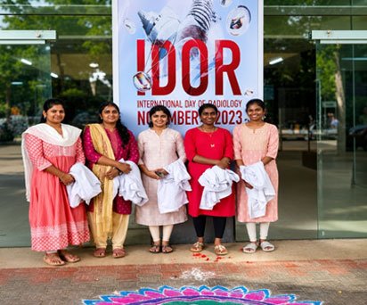
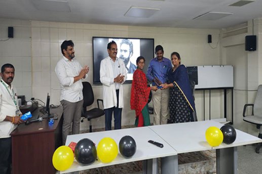
The Department of Radiodiagnosis proudly celebrated the International Day of Radiology (IDOR) on 8 November 2023.
CME / Workshops / Conferences
Undergraduate Course (MBBS)
15 days OP posting during 2nd clinical year and theory classes as per revised teaching schedule are taken to the prefinal year students.
The Department of Radiodiagnosis provides structured academic teaching and clinical exposure for MBBS undergraduate students as part of their medical curriculum.
Undergraduate (MBBS) Teaching Programme
The Department of Radiodiagnosis provides structured academic teaching and clinical exposure for undergraduate medical students as per the curriculum guidelines.
During the 2nd Clinical Year, students undergo a 15-day clinical posting in the Department of Radiodiagnosis. In addition, theory classes are conducted for MBBS Pre-Final Year students as per the revised teaching schedule.
Theory classes are conducted for Pre-Final Year MBBS students.
Classes are conducted every Tuesday from 3:00 PM to 4:00 PM.
The teaching programme is conducted during the second half of the academic year (July to December).
Students are provided with knowledge of basic radiological principles, imaging modalities, radiation safety, and interpretation of common radiological findings.
The undergraduate teaching programme aims to provide students with fundamental knowledge of diagnostic imaging modalities, interpretation of common radiological findings, radiation safety, contrast procedures, and principles of modern imaging techniques.
UG (MBBS Pre-Final Year) Theory Class Schedule
| S.No | Topic |
|---|---|
| 1. | Basic Principles of X-Ray and Radiation Safety Measures |
| 2. | Fluoroscopy / IITV – Basic Principles |
| 3. | Chest X-Ray – Basics and Interpretation |
| 4. | Abdominal X-Ray – Basics and Interpretation |
| 5. | X-Ray Contrast Procedures: Barium Swallow, Barium Meal, Barium Enema, Loopogram |
| 6. | X-Ray Contrast Procedures: IVU, AUG, MCU, PCN, Fistulogram, Sinogram, Sialogram |
| 7. | Ultrasound Basics, Patient Preparation and Indications |
| 8. | Focused Ultrasound in Emergency Medicine |
| 9. | Obstetric Ultrasound – Basic Principles |
| 10. | Doppler Ultrasound – Basic Principles |
| 11. | Computed Tomography (CT) – Basic Principles |
| 12. | CT Brain – Anatomy and Common Pathologies |
| 13. | CT Abdomen – Patient Preparation, Scan Protocols and Anatomy |
| 14. | CT Multiplanar Reconstruction (MPR) and CT Angiography |
| 15. | MRI Basics, Sequences and Safety Measures |
| 16. | MRI Brain – Basic Anatomy and Pathology |
| 17. | MRI Abdomen and Pelvis – Basic Principles |
| 18. | Ultrasound of Small Parts – Basic Principles |
| 19. | Contrast Media – Types, Uses and Safety |
| 20. | Nuclear Medicine – Radioactivity, Radiotracers and Safety Measures |
| 21. | Nuclear Medicine Studies: Ventilation–Perfusion Scan and Bone Scan |
| 22. | Radiation Dose, Biological Effects, Risks and Safety Measures |
| 23. | Anatomical and Functional Imaging, PET and Fusion Imaging |
| 24. | Contrast Media Reactions and Management |
Teaching Methodology
- Classroom-based theory sessions
- Clinical case discussions
- Demonstration of imaging modalities
- Exposure to X-ray, Ultrasound, CT, MRI, Nuclear Medicine, and PET imaging
- Practical understanding of radiation protection and patient safety
Learning Outcomes
After completion of the posting, undergraduate students will be able to:
- Understand the principles of various radiological imaging techniques
- Identify common radiological findings
- Understand appropriate indications for imaging investigations
- Learn basic interpretation of X-ray, CT, MRI, and Ultrasound studies
- Understand radiation hazards and safety practices
- Gain awareness of contrast media usage and adverse reactions
Paramedical Course – Diploma in Radiodiagnostic Technology (DRDT)
Department of Radiodiagnosis, Government Mohan Kumaramangalam Medical College Hospital (GMKMCH), Salem
The Department of Radiodiagnosis conducts the Diploma in Radiodiagnostic Technology (DRDT) course to train students in the field of medical imaging technology. The course provides comprehensive theoretical knowledge and practical training in modern radiological imaging techniques.
Course Details
| Course Name | Details |
|---|---|
| Course Name | Diploma in Radiodiagnostic Technology (DRDT) |
| Duration | 2 Years |
| Annual Intake | 10 Students per Year |
| Affiliation | Tamil Nadu Dr. M.G.R. Medical University, Chennai |
Course Objectives
The DRDT course aims to develop skilled radiological technologists with knowledge and practical expertise in diagnostic imaging procedures, radiation safety, patient care, and equipment handling.
Curriculum & Training Areas
- Radiological Imaging Techniques
- Contrast Imaging Procedures
- Pharmacology & Patient Care
- MRI Training
Student Assessment
Student performance is evaluated through:
- Periodic theory examinations conducted every fortnight
- Practical training assessment
- Clinical case discussions
- Departmental academic activities
Training Highlights
- Hands-on exposure to advanced imaging modalities
- Training under experienced Radiologists and Technical Staff
- Clinical exposure in X-ray, CT, MRI, Ultrasound, and Fluoroscopy services
- Practical experience in patient positioning, imaging protocols, and departmental workflow
Research Papers and Publications
| S.No | Faculty Name | Details of Publication |
|---|---|---|
| 1. | Dr. P. Kumar MDRD., | Kumar P, Arun C. MRI colonography versus conventional colonoscopy in detection of colonic polyposis. International journal of latest research in science and technology, March–April 2016; volume (5) Issue (2): page number 80-85. Available from: mnkjournals.com/ijlrst.htm |
| Arun C, Kumar P. Utility of Transrectal ultrasound with power Doppler imaging guided biopsy in the detection of prostate cancer. International journal of latest research in science and technology, March–April 2016; volume (5) Issue (2): page number 40-43. Available from: mnkpublication.com/journal/ijlrst/index.php | ||
| Kumar P, Arun C. MRI Evaluation of ligamentous injuries and meniscal injuries of the knee joint. International journal of science and research, February 2019; volume (8) Issue (2): page number 2245-2249. Available from: ijsr.net | ||
| Kumar P, Arun C. Evaluation of Fistula in Ano MR Fistulography. International journal of science and research, February 2019; volume (8) Issue (2): page number 2250-2254. Available from: ijsr.net | ||
| 2. | Dr. N. Sundareswaran MDRD., | Sundareswaran N, Babu Peter, Kailasanathan. In phase opposed phase sequence in MRI differentiating neoplastic lesions of bone marrow from non-neoplastic lesions of bone marrow. International journal of latest research in science and technology, January-February 2016; volume (5) Issue (1): page number 146-156. Available from: mnkjournals.com/ijlrst.htm |
| Shankaranandh R, Sundareswaran N. Characterization of Adrenal masses with contrast enhanced CT-washout study. International journal of latest research in science and technology, May-June 2016; volume (5) Issue (3): page number 6-10. Available from: mnkjournals.com/ijlrst.htm | ||
| Kalaivani P, Sundareswaran N. Hippocampal abnormalities in Adults with unilateral temporal lobe Epilepsy - A Diffusion Tensor imaging study. International journal of science and research, November 2019; volume (8) Issue (11): page number 1564-1571. Available from: ijsr.net | ||
| Sundareswaran N, Elanchezhian E. Role of time intensity curve in dynamic contrast MRI evaluation of soft tissue tumour. International journal of contemporary medicine surgery and Radiology, April-June 2020; volume (5) Issue (2): page number B60-B65. Available from: dx.doi.org/10.21276/ijcmsr.2020.5.2.14 | ||
| 3. | Dr. M. Sankar MDRD., | Sankar M, Vidhya E. Descriptive study on various perianal fistulas in rural population using MR Fistulogram. International journal of Radiology and Diagnostic Imaging, February-2021; volume (4) Issue (1): page number 228-230. Available from: dx.doi.org/10.33545/26644436.2021.v4.i1d.189 |
| Vidhya E, Sankar M. Correlation of CT pornography vasculature to child Pugh classification of cirrhotic patients. International journal of Radiology and Diagnostic Imaging, February-2021; volume (4) Issue (1): page number 231-233. Available from:dx.doi.org/10.33545/26644436.2021.v4.i1d.190 | ||
| 4. | Dr. E. Vidhya DMRD., DNB., | Sankar M, Vidhya E. Descriptive study on various perianal fistulas in rural population using MR Fistulogram. International journal of Radiology and Diagnostic Imaging, February-2021; volume (4) Issue (1): page number 228-230. Available from:dx.doi.org/10.33545/26644436.2021.v4.i1d.189 |
| Vidhya E, Sankar M. Correlation of CT pornography vasculature to child Pugh classification of cirrhotic patients. International journal of Radiology and Diagnostic Imaging, February-2021; volume (4) Issue (1): page number 231-233. Available from:dx.doi.org/10.33545/26644436.2021.v4.i1d.190 |
Department Census Details
Department of Radiodiagnosis
The Department of Radiodiagnosis provides comprehensive diagnostic imaging services to outpatients, inpatients, emergency cases, and referred patients. The department maintains detailed records of imaging workload and service utilization across various sections.
Annual Imaging Workload Summary
The department provides advanced diagnostic services including:
- Digital X-Ray and Mammography
- Computed Tomography (CT) and CT Angiography
- Magnetic Resonance Imaging (MRI)
- Ultrasound and Colour Doppler Studies
- Contrast Radiological Procedures
- Image-guided Interventional Procedures
Major Service Volume Highlights
| Year | Total X-Ray Cases | CT Scans | MRI Scans | USG Studies |
|---|---|---|---|---|
| 2020 | 68,419 | 53,252 | 4,116 | 28,613 |
| 2021 | 66,522 | 80,065 | 5,769 | 36,256 |
| 2022 | 1,05,465 | 69,400 | 8,118 | 53,676 |
| 2023 | 1,15,637 | 69,729 | 9,236 | 70,869 |
| 2024 | 1,06,085 | 69,167 | 10,686 | 95,669 |
| 2025 | 1,10,000 | 72,000 | 11,000 | 98,000 |
COVID-19
During the COVID-19 pandemic, the Department of Radiodiagnosis played a vital role in the diagnosis, assessment of disease severity, and management of COVID-19 patients through CT imaging.
Highlights
- Dedicated CT services supported rapid assessment of COVID-19-related lung involvement.
- The department provided continuous imaging support during the peak pandemic period.
- CT imaging contributed significantly to patient triaging, treatment planning, and follow-up evaluation.
First Batch of Postgraduate Students Successfully Graduates – 2025
The MD Radiodiagnosis postgraduate degree programme was established in the academic year 2022–2023 with an approved annual intake of three students. The programme was initiated with the objective of training competent, ethical, and skilled radiologists capable of meeting the evolving demands of modern healthcare.
The Department of Radiodiagnosis at Government Mohan Kumaramangalam Medical College Hospital (GMKMCH), Salem, proudly announces the successful graduation of its first batch of Postgraduate (MD Radiodiagnosis) students in 2025. This landmark achievement marks a significant milestone in the department's academic journey and reflects its continued commitment to excellence in postgraduate medical education, clinical training, research, and patient care.
Since the inception of its postgraduate program, the department has been dedicated to providing comprehensive training in diagnostic imaging through experienced faculty, modern imaging facilities, and a strong emphasis on academic excellence and ethical medical practice.
The successful completion of training by the inaugural batch stands as a testament to the department's sustained efforts in nurturing competent radiologists who are well-equipped to contribute to healthcare, medical education, and research. The Department extends its heartfelt congratulations to the graduating students and wishes them continued success in their professional careers.
This achievement reinforces the Department of Radiodiagnosis at GMKMCH, Salem, as a center of excellence in radiology education and patient care, paving the way for many more successful batches in the years to come.
PGS Students Excel at IRIA Meet Quiz Competition
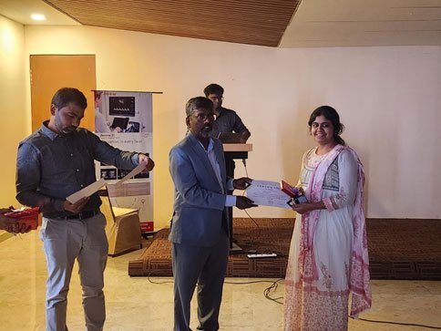
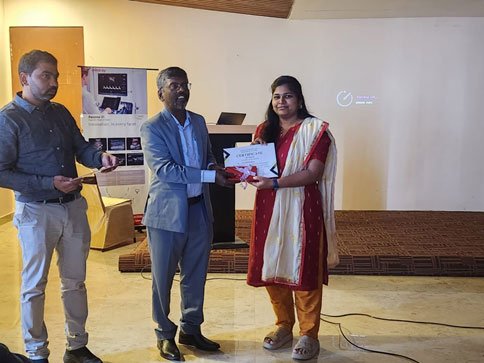
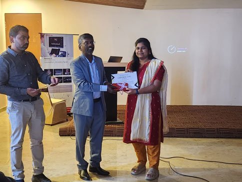
PGS students secured First and Second Prizes in the Quiz Competition held during the IRIA Subchapter Meet at Yercaud, bringing pride to the institution with their outstanding performance. Congratulations to the winners!
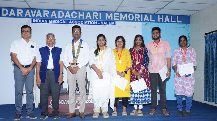
PGS Students Win at IMA Salem District-Level Quiz Competition
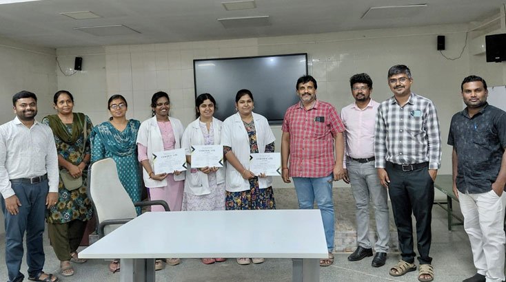
PGS students secured First Prize in the District-Level Quiz Competition organized by the Indian Medical Association (IMA), Salem. Their outstanding performance reflects the institution's commitment to academic excellence. Congratulations to the winners!
Special Interventional Radiology Program
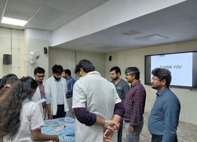
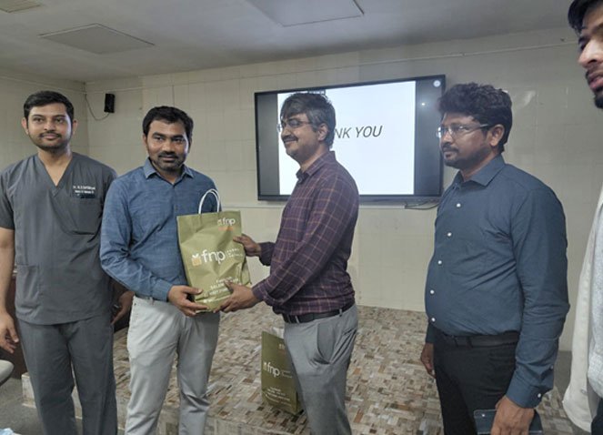
The Department of Radiology successfully organized a Special Interventional Radiology Program featuring an insightful guest lecture by consultant interventional radiologists, Dr. P. Santhosh Kumar and Dr. Karthikeyan. The session highlighted recent advances, innovative image-guided procedures, and the expanding role of Interventional Radiology in modern clinical practice. The program was well received by faculty members, postgraduate residents, and students, providing an excellent platform for academic learning and professional interaction.