
Introduction
History of the Department
Government Mohan Kumaramangalam Medical College, the prestigious institution of the Steel City, was started on 23rd April 1986 by upgrading the existing District Government Hospital which was started in 1917 in a span of 150 acres of land, in order to serve the sick and needy people of Salem and the adjacent districts. The foundation for the Government Mohan Kumaramangalam Medical College was laid at Salem by the late Shri. Rajiv Gandhi, the then Prime Minister of India, on 25th October 1988. Admission of students for the first batch was carried out in July 1990. The Non-Teaching Headquarters Hospital, Salem was converted into the teaching Government Mohan Kumaramangalam Medical College Hospital, which is now well equipped with all modern equipment and qualified staff.
The College, which was temporarily situated in the District Board Building, was moved to this 150-acre premises on the Steel Plant Road. The majestic faculty block has enormous space for the various departments and their museums. The College has a separate lecture hall block, administrative block, animal house and a spacious library block. A big modern auditorium and two larger lecture halls with the latest audio-visual equipment were inaugurated in April 2005. This College has been recognized for the award of the M.B.B.S. degree granted by the Tamilnadu Dr. M.G.R. Medical University, vide letter no. MCI 34(A1)/96/Med/8661 dated 8/7/96 of the Medical Council of India, New Delhi.
Department of Anatomy was innovated in 1991 with qualified and experienced faculty members with a research background. Innovative learning and teaching methodologies have been adopted for future medical and para-medical professionals. With the growth of the institute, the department has expanded to impart undergraduate training and other courses such as BSc Nursing and Allied Medical Sciences. We seek to instill in our students a sense of wonder at the beauty of the human body, and hope that our students learn values of respect for the cadaver, themselves and others during their time here. The students spend their first year of training in the basic science departments, nearly 50% of which time is spent in the Anatomy department, where they are grounded thoroughly in Gross Anatomy, Histology, Embryology, Osteology and Genetics with special emphasis on Clinical Anatomy. The gross structure of the human body is taught through lectures, cadaver dissections, study of bones, prosected specimens, cross-sections, tutorials, quizzes, radiographs, charts and models. Even though the students do not have to dissect as a part of their practical exams, learning throughout the year is facilitated by dissection performed in groups of 10–15 to ensure that all students receive personal attention and guidance. Microscopic anatomy is taught through lectures and practical sessions. The Histology Laboratory is well equipped with individual microscopes.
Teaching Faculties
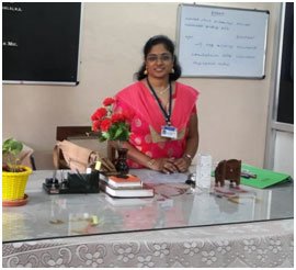
Dr. Kalai. Anbusudar, MD.,
Professor & HOD
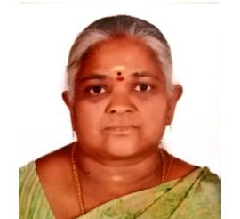
Dr. Kalai. Anbusudar, M.D., Professor of Anatomy, has been conducting various academic activities for the past 10 years for MBBS and para-medical students like quiz programs, guest lectures, embryology and histology model competitions, integrated teaching programs and mentoring. She has published articles in various indexed National and International journals. She has completed the Revised Basic Course workshop in Medical Education at JIPMER, Puducherry, and the Advance Course in Medical Education at CMC, Vellore. She has undergone the Curriculum Implementation Support Programme and Basic Course in Biomedical Research. An ICMR project has also been conducted under her co-guidance. She has been appointed as external examiner for MBBS and paramedical university examinations. She has received the Best Doctor Award and appreciation certificate from the Dean, GMKMC, Salem‑30.
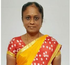
Dr. M.R. Manimekalai, M.D, DCH
Associate Professor
Dr. M.R. Manimekalai, M.D., DCH, Associate Professor of Anatomy, has been conducting various academic activities for MBBS and para-medical students like quiz programs, guest lectures, embryology and histology model competitions, integrated teaching programs and mentoring. She has published articles in various indexed National and International journals. She has undergone the Revised Basic Course in Medical Education Technologies, Curriculum Implementation Support Programme and Basic Course in Biomedical Research.
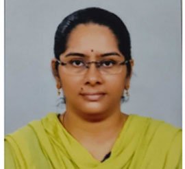
Dr. K. Aruna, M.D, DCP
Senior Assistant Professor
Dr. K. Aruna, M.D., DCP, Senior Assistant Professor in Anatomy, has been conducting various academic activities for MBBS and para-medical students like quiz programs, guest lectures, embryology and histology model competitions, integrated teaching programs and mentoring. She has undergone the Revised Basic Course in Medical Education Technologies, Curriculum Implementation Support Programme and Basic Course in Biomedical Research. She has been appointed as external examiner for MBBS and paramedical university examinations. She has received the Best Doctor Award and appreciation certificate from the Dean, GMKMC, Salem‑30.
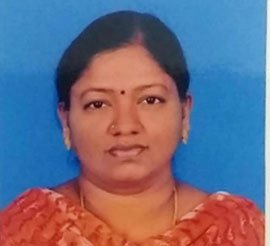
Dr. C. Sasikala, M.D, DCP
Senior Assistant Professor
Dr. C. Sasikala, M.D., DCP, Senior Assistant Professor in Anatomy, has been conducting various academic activities for MBBS and para-medical students like quiz programs, guest lectures, embryology and histology model competitions, integrated teaching programs and mentoring. She has published articles in various indexed National and International journals. She has undergone the Revised Basic Course in Medical Education Technologies and Basic Course in Biomedical Research.
Tutors
- Dr. Rathinavel
- Dr. Manikam Kalaiselvi
- Mrs. P. Vennila, M.Sc
Infrastructure
Dissection Hall
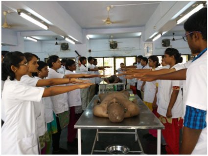
Cadaveric dissection is an integral part of medical education and the basis of anatomy for first M.B.B.S. students. Dissection of cadavers is a time-honoured part of medical education. Undergraduates do dissection as a part of learning human anatomy. Cadavers are obtained from the forensic mortuary declared as an ‘unclaimed body’, or through donated bodies that include voluntary donation during life or after demise by their relatives.
The dissection hall is well equipped according to MCI guidelines. It is well lit and well ventilated with exhaust fans. There is a tank room in the dissection hall with adequate preservation tanks for cadavers and dissected specimens. There is an ante-room for students with lockers and wash basins with water taps. The dissection hall is well equipped with dissection tables, handsaw, cutting machine, X-ray view box, instruments, equipment & wall charts.
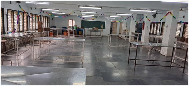
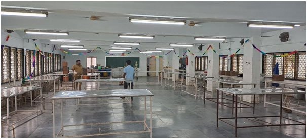
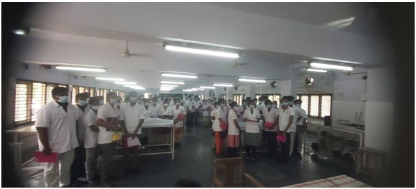
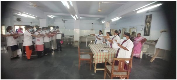
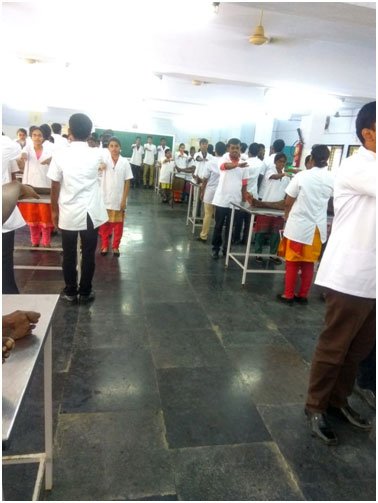
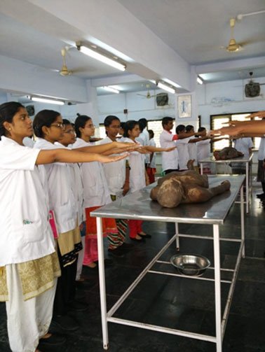
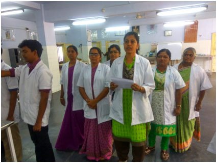
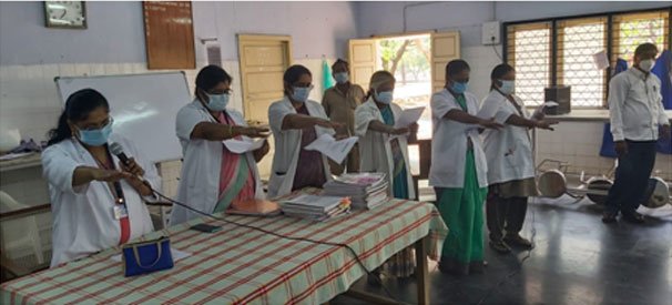
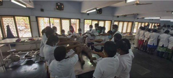
Cadaveric oath is taken every year while starting dissection for first year MBBS students.
Embalming Room
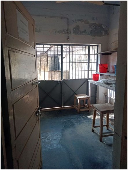
Embalming room is available adjacent to the dissection hall, with adequate electrical embalming machines.
Bone Room
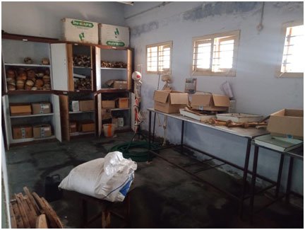
There is a bone room in the dissection hall, with an adequate number of articulated and disarticulated bones necessary for the students.
Burial Ground
A burial ground is present close to the dissection hall, according to MCI norms, for disposal of decomposed cadavers and specimens. Bones are collected periodically from the pits for study purposes.
Cold Storage Room
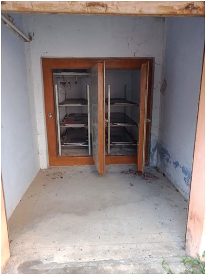
One cold storage room is available with adequate space and uninterrupted power supply.
Museum
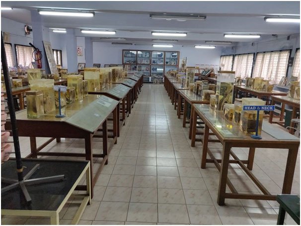
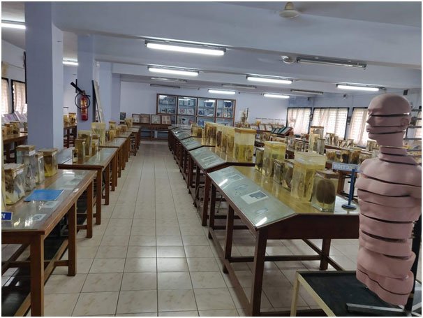
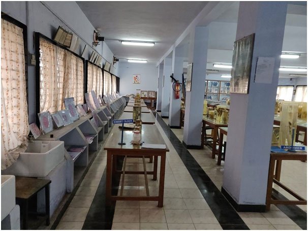
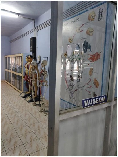
A museum displaying a number of instructive and explicit gross anatomy and embryology specimens, preserved and mounted with dexterity, and many models which have been catalogued according to regions. These silent teachers (preserved human specimens) are always there, enabling students to enrich their knowledge in Anatomy. The museum is well provided with adequate seating accommodation.
Histology Lab
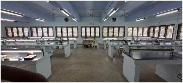
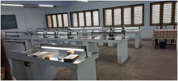
The histology laboratory is fully fledged with all the basic facilities, equipped with enough light microscopes and dissection microscopes for research and teaching purposes. There are a number of laminated histology charts for the students to get a clear understanding of microanatomy.
Histology Research Lab
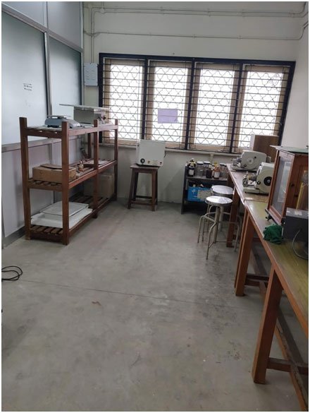
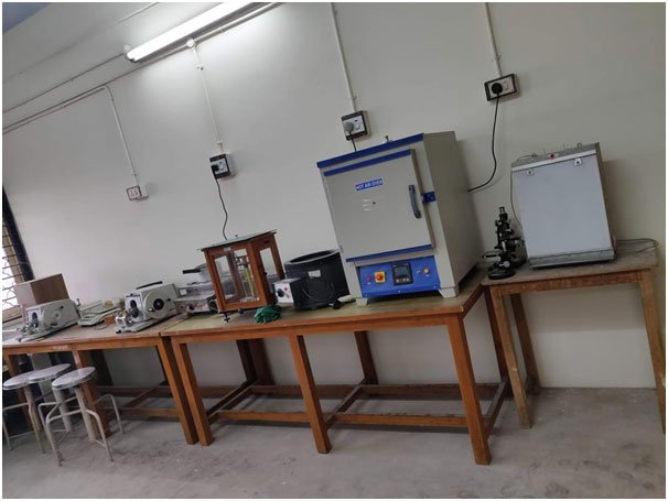
The research laboratory is well equipped with binocular research microscopes, stereoscopic dissecting microscope, incubator, hot plate, rotary microtome, microtome knife, etc. The research laboratory provides an ideal environment for the faculty and students to carry out research activities.
Library
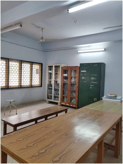
The department has a library with journals & books covering various sub-sections of anatomy, including gross anatomy, histology, embryology, neuroanatomy, osteology, surface markings, embalming and radiological anatomy.
Demonstration Room
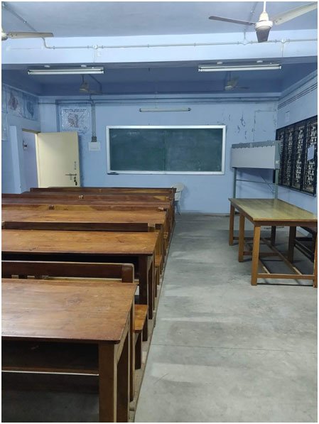
Two demonstration rooms, each of 60 sq. m, accommodating 75 students and equipped with appropriate audio-visual aids, add to the teaching ambience of the department.
Lecture Hall
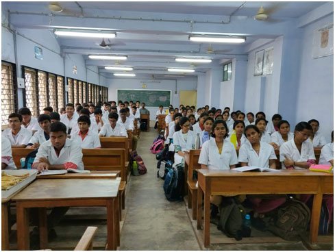
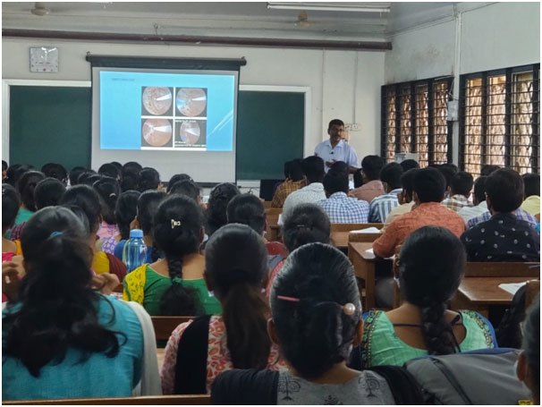
Six well-furnished and spacious lecture halls, each with a seating capacity of 200. A well-furnished auditorium with a seating capacity of 325, and a specially built exam hall for 300 students. Halls equipped with the latest audio-visual equipment like OHPs, LCD projectors, computers, cordless microphones, etc.
Office Room
An office room for lab technicians and maintenance of official records is available.
Faculty Room
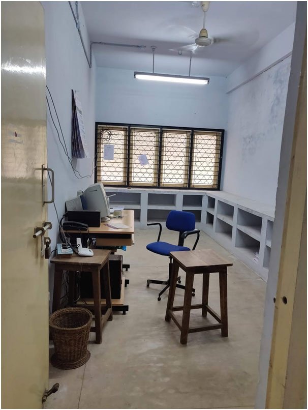
Adequate faculty rooms are available for HOD, Associate Professors, Assistant Professors and Tutors.
Store Room
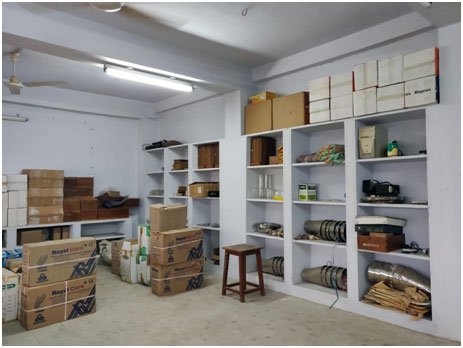
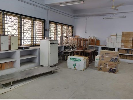
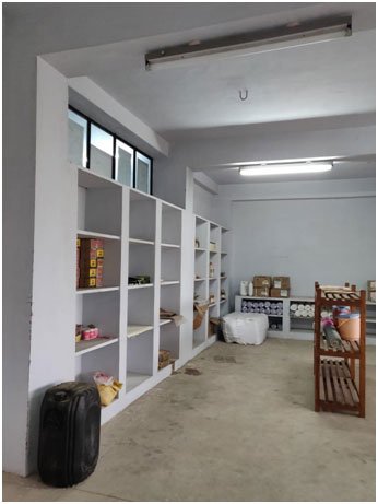
Store rooms are present for maintenance of chemicals, equipment and furniture.
Voluntary Body Donation
The forms of willingness of volunteers who intend to donate their body after death to the department are received, and after death, cadavers are brought to the department by relatives.
Academic Activities
UG Courses
- MBBS
- B.Sc. Nursing
- B.Sc. Allied Medical Courses
- B.Sc. (MLT)
- B.Sc. (Operation Theatre Technology)
- B.Sc. (Cardiac Technology)
- B.Sc. (Radio-therapy Technology)
Periodic Academic Activities
- Didactic lectures
- Dissections
- Pre-dissection classes
- Demonstrations
- Histology practicals
- Clinical anatomy (OSPE charts)
- Symposiums
- Integrated teaching, both horizontal and vertical
- Daily slip test of previous classes
- Internal assessment test, both theory and practical
- Record work — both macroscopic and microscopic anatomy
- Daily practice of diagrams
- Radiological anatomy (basics in X-rays, CT, MRI)
- Developmental anatomy (lectures, models, charts)
- Osteology classes (articulated skeleton, disarticulated bones)
- Surface anatomy classes
- Early clinical exposure
- Self-directed learning
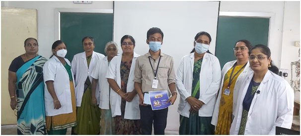
- Quiz programs
- Model competitions
- Hands-on workshops for UGs and PGs
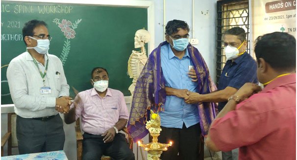
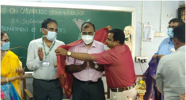
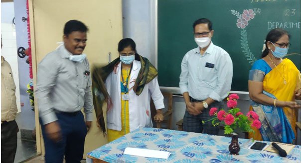
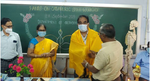
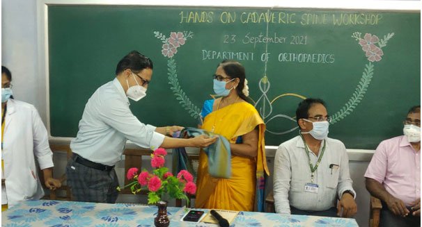
- Hands-on workshop on cadaveric dissection by Department of Anatomy and Department of Orthopaedics
Other Academic Activities
Intra-Departmental Quiz
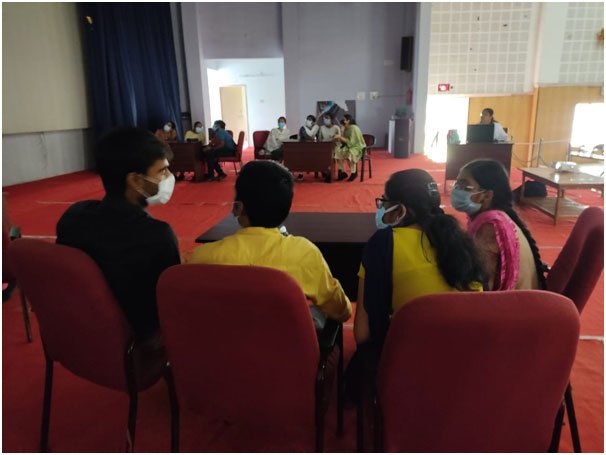
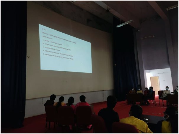
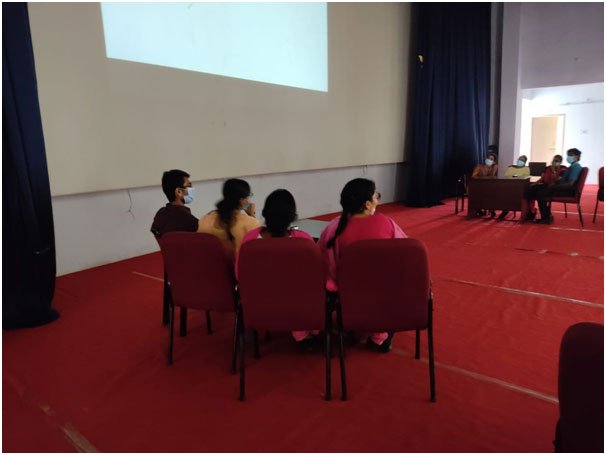
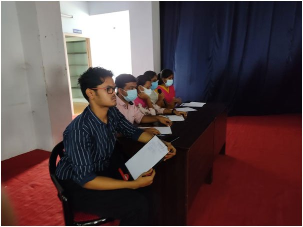
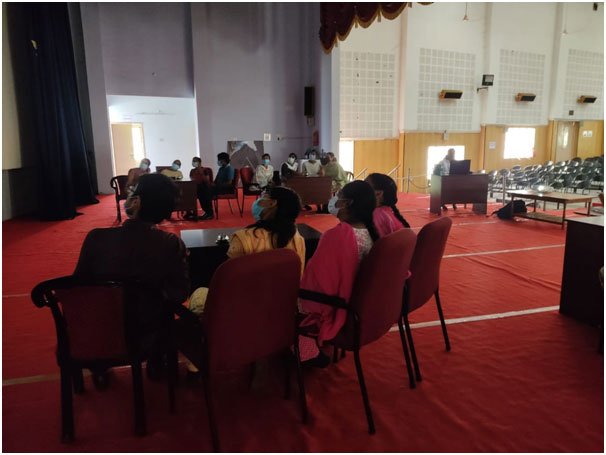
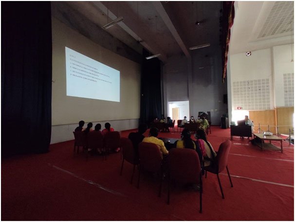
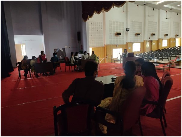
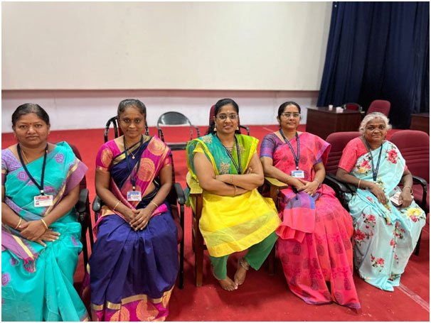
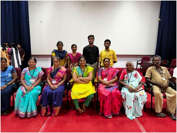
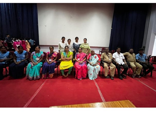
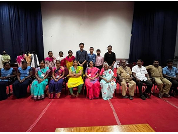
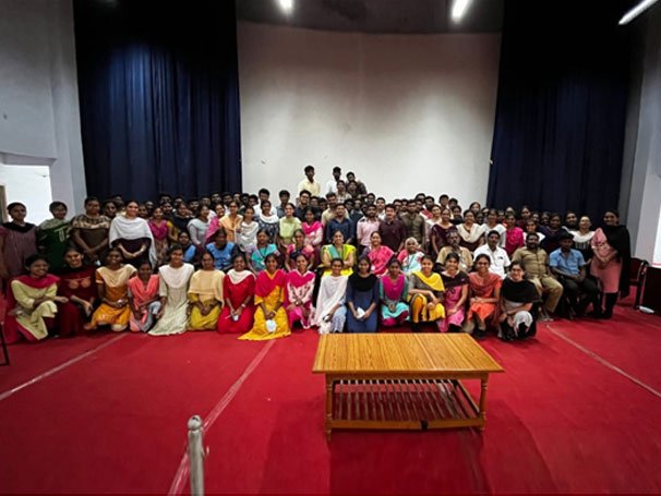
Every year the Department of Anatomy conducts intra-departmental quiz programmes for first year MBBS students. Prizes are awarded to the winners and to those who answer correctly among the audience.
Early Clinical Exposure
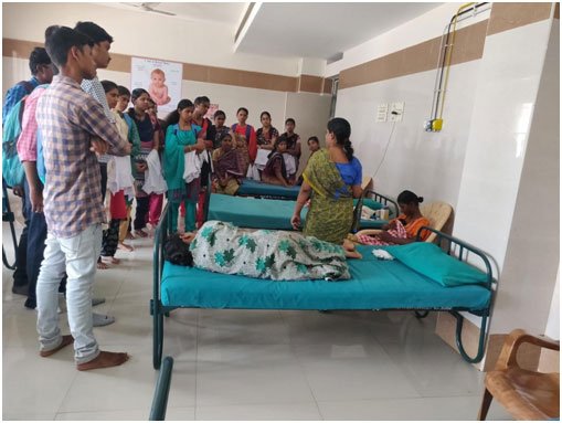
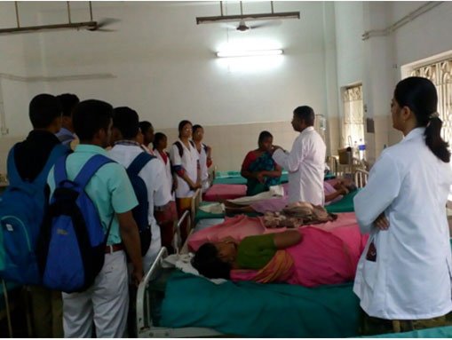
Based on the medical education curriculum for first MBBS students, for early clinical exposure, students are taken to various clinical departments (viz. General Medicine, General Surgery, Obstetrics and Gynaecology, Orthopaedic Surgery and Paediatric Medicine).
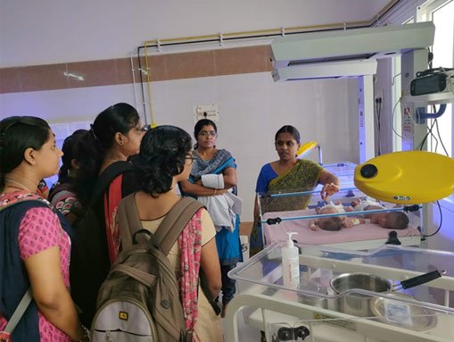
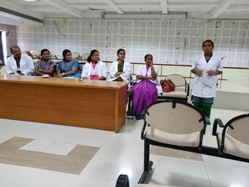
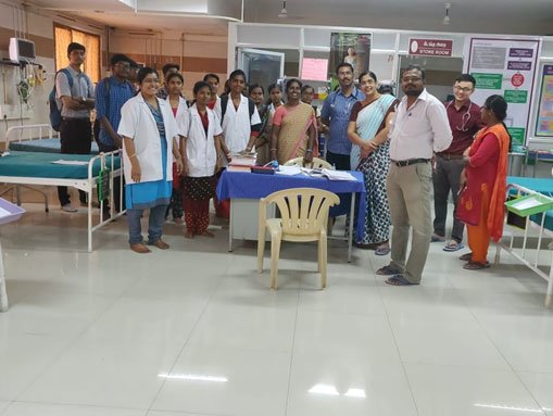
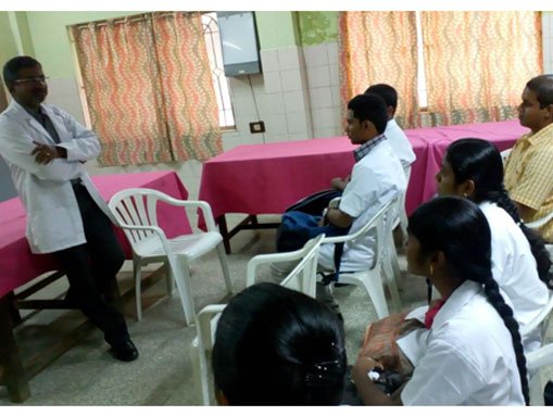
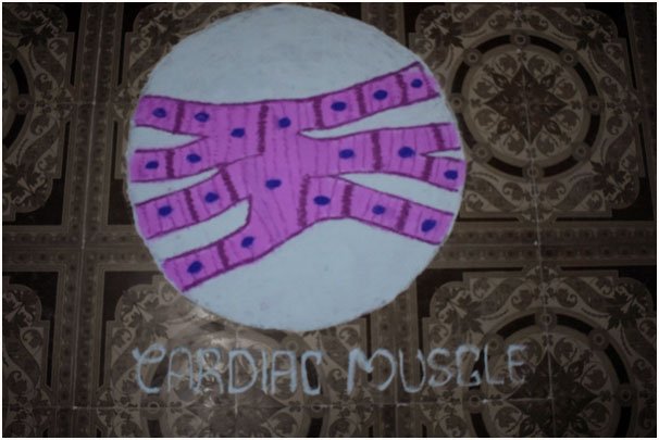
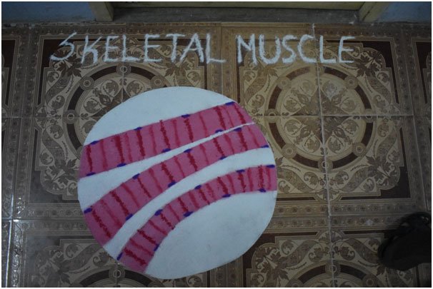
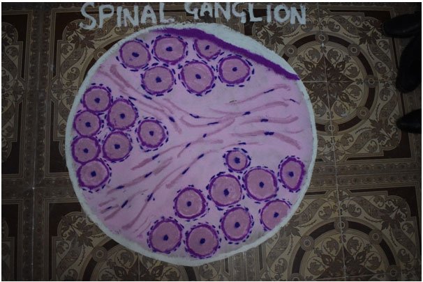
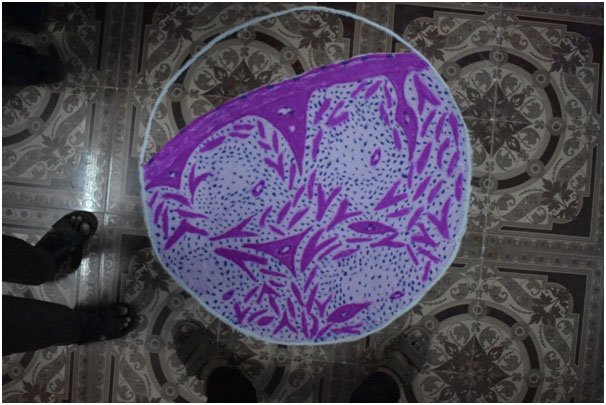
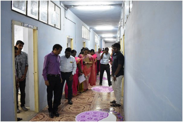
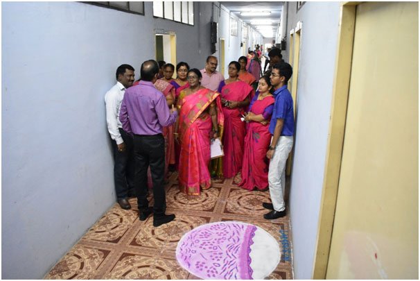
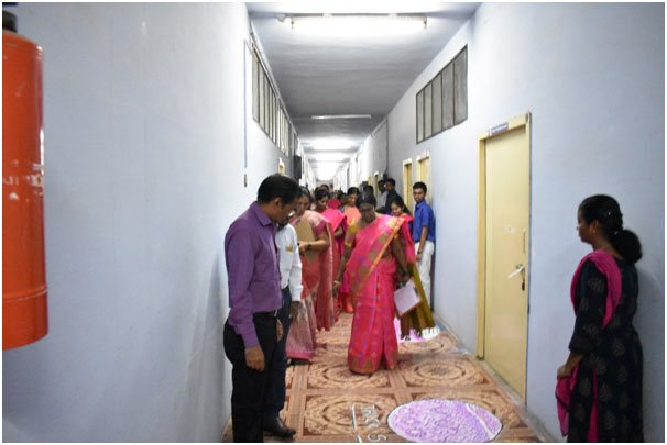
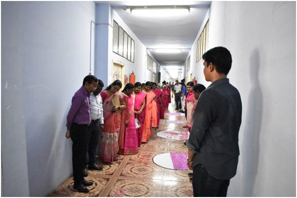
Histology Rangoli Competition — I MBBS Students, 2017 Batch
Guest Lecture with Embryology Model Competition
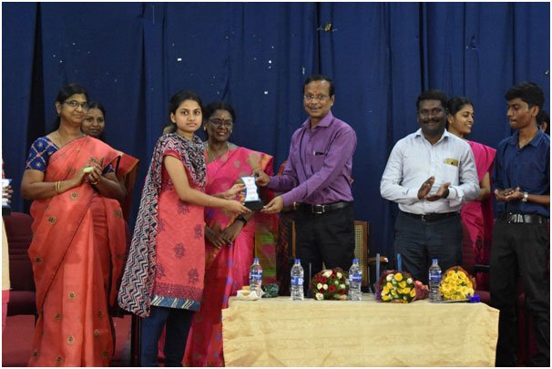
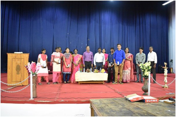
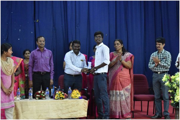
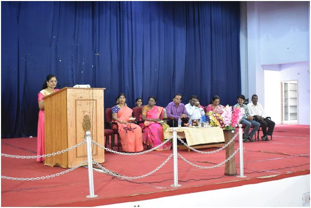
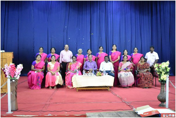
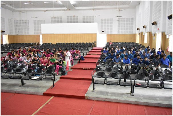
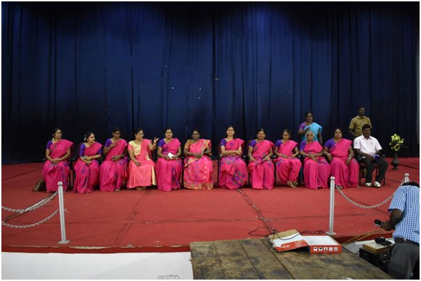
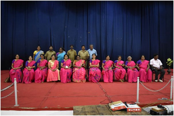
As a part of academic activity, a guest lecture was conducted along with an Embryology Model Competition for the first MBBS 2016–17 batch students. The topic was “The Brain Stem and Its Clinical Aspects” by Prof. Dr. Sheela Grace, M.S. The best models were awarded prizes as encouragement for students and to stress the importance of Embryology.
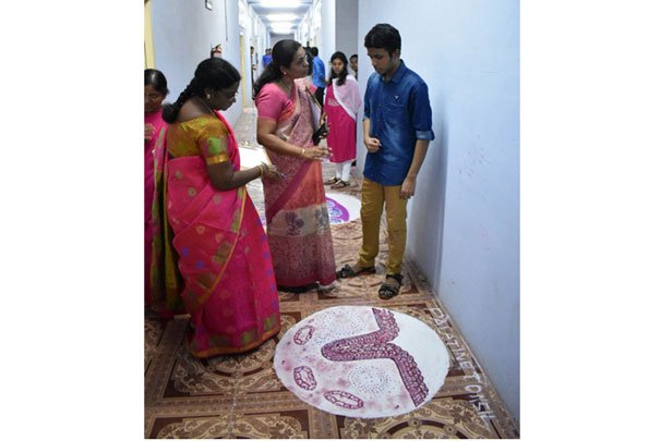
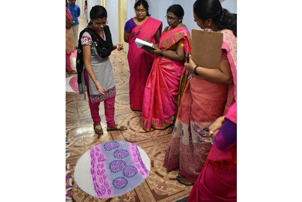
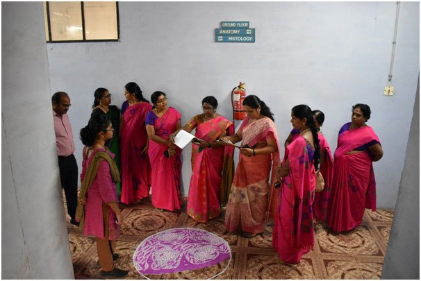
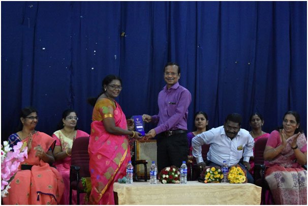
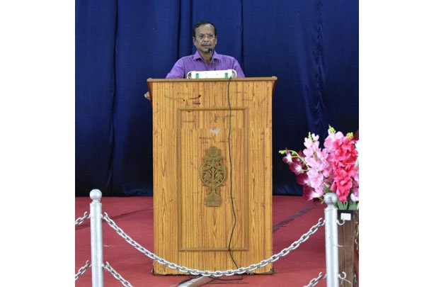
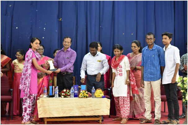
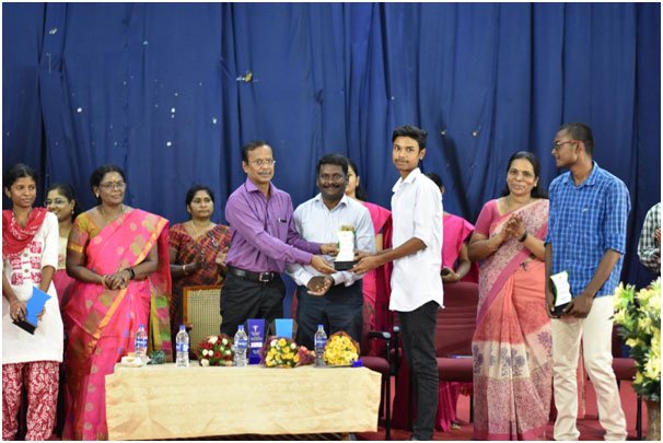
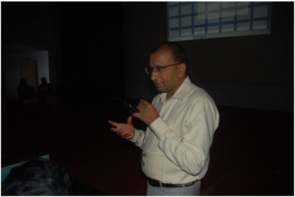
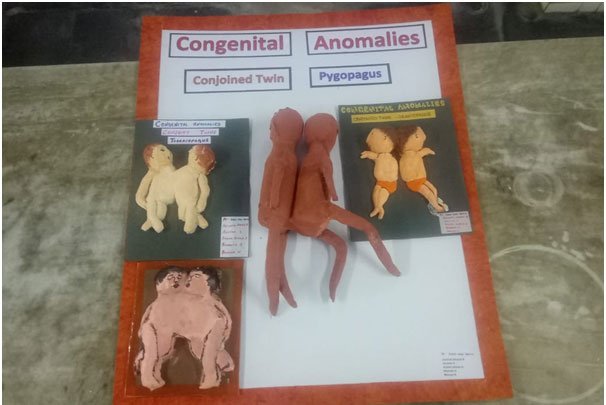
Guest Lecture with Congenital Anomalies Model Competition — 2020 MBBS Batch
Dr. S. Dhivya, M.D., Professor and HOD of Anatomy, Government Namakkal Medical College & Dr. Pachiyappan, M.D., D.M, Cardiologist, GMKMCH, Salem
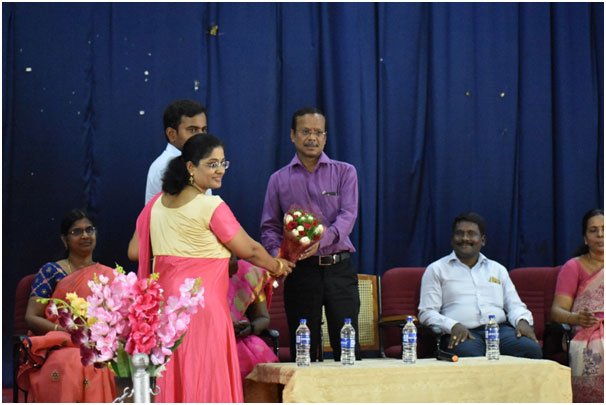
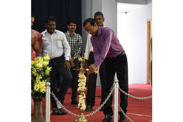
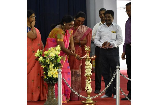
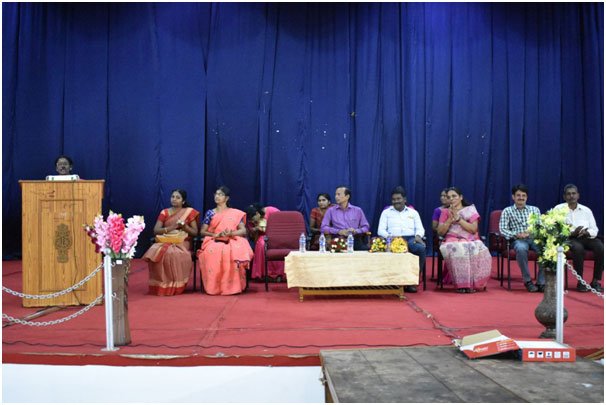
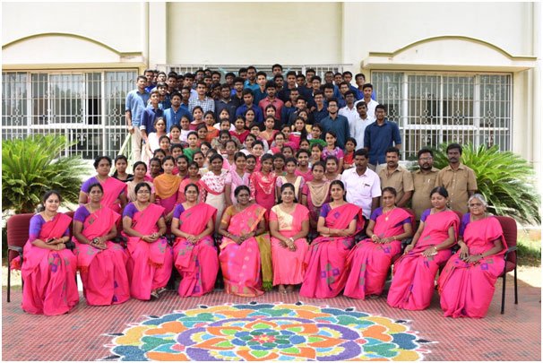
Guest Lecture — Additional Session
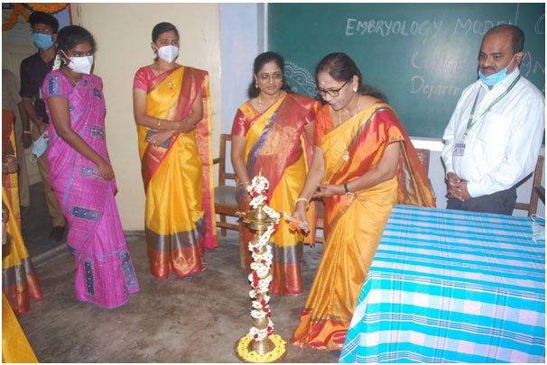
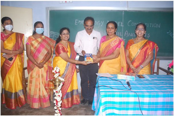
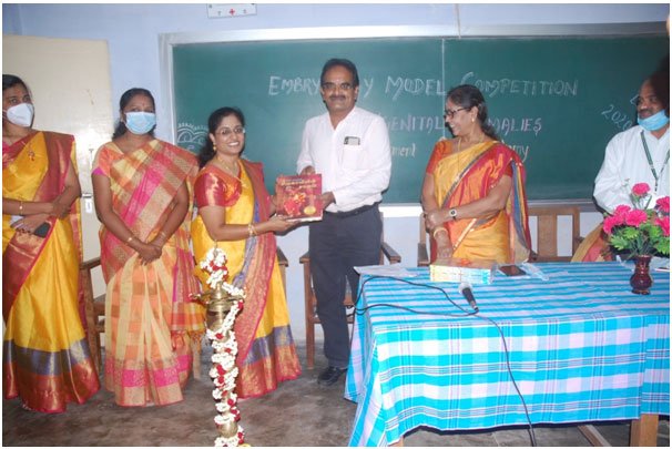
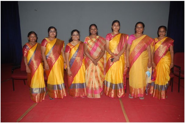
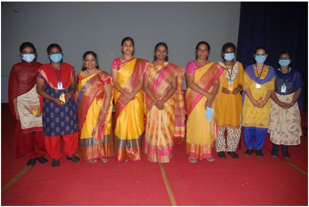
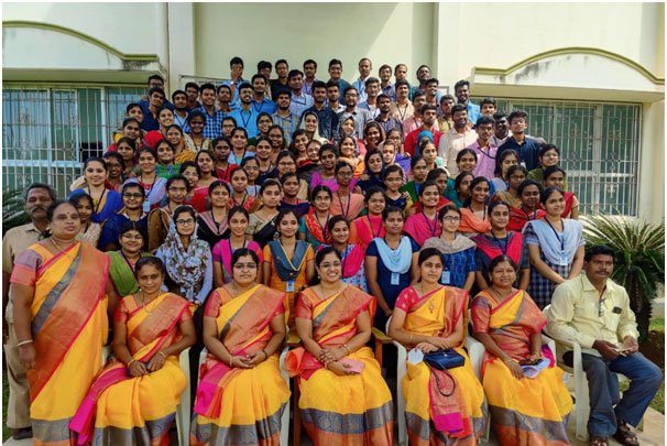
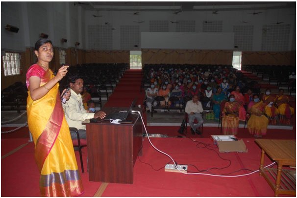
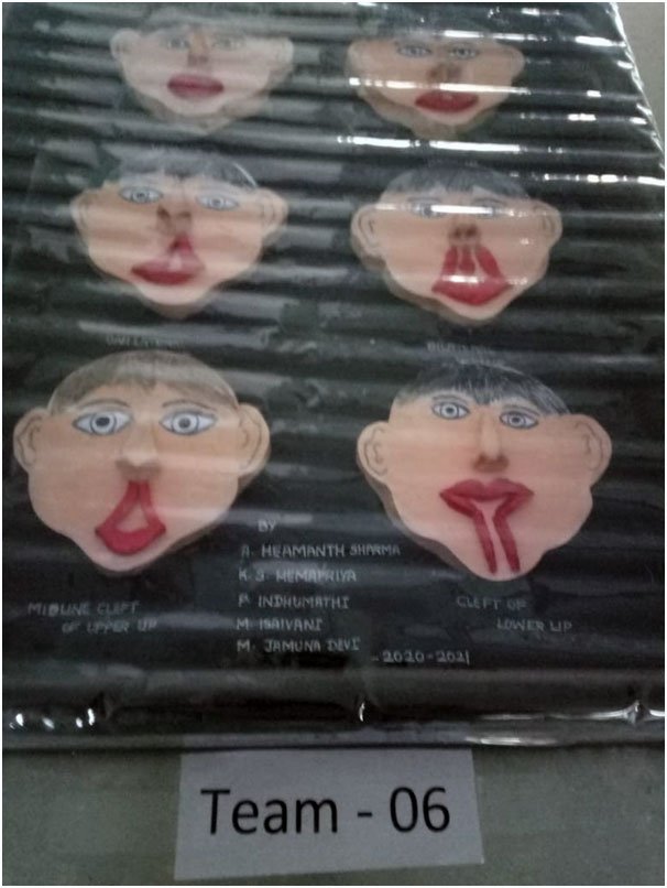
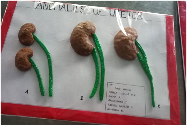
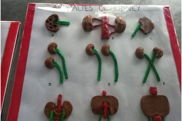
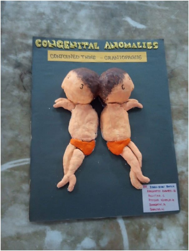
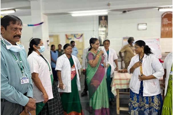
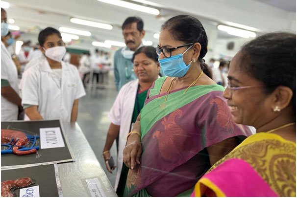
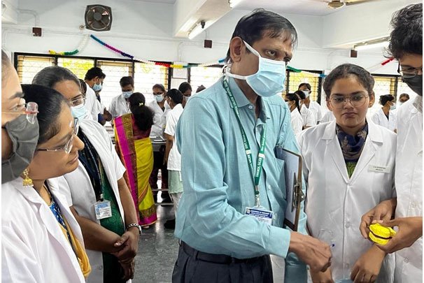
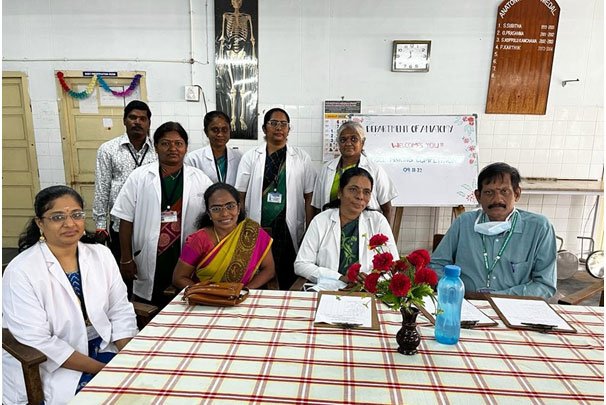
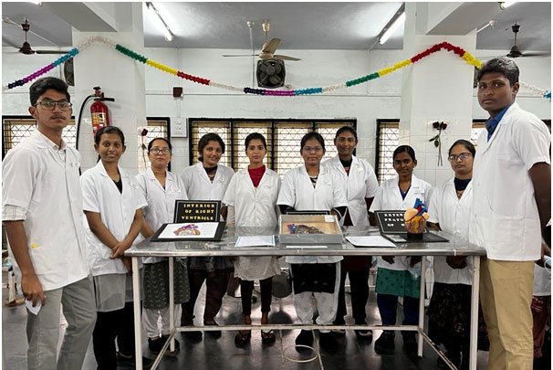
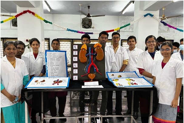
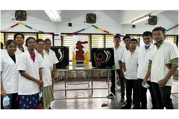
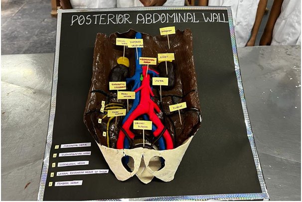
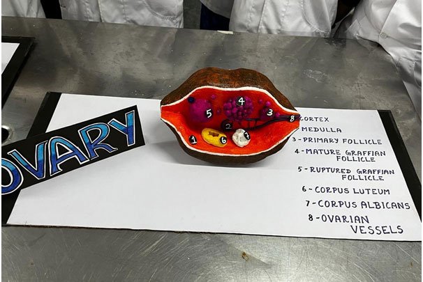
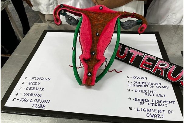
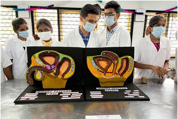
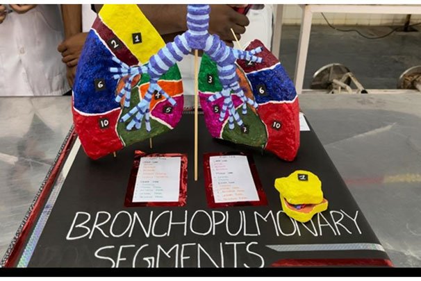
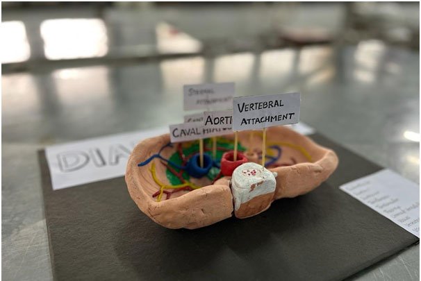
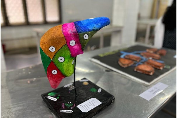
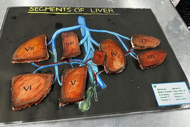
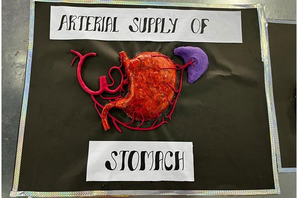
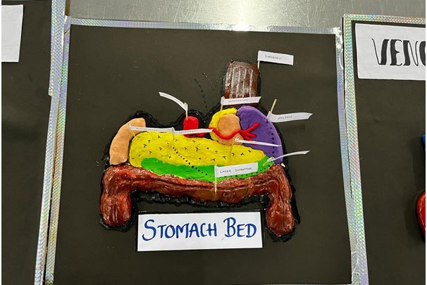
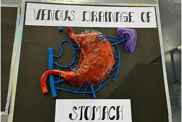
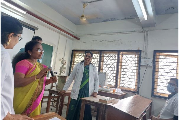
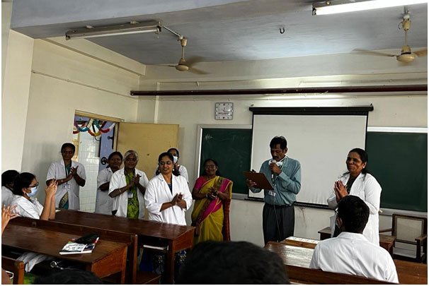
Gross Anatomy Models Competition — 2021 Batch MBBS Students
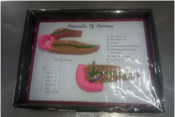
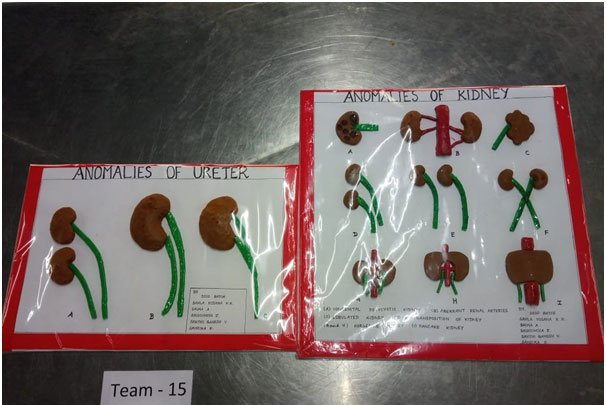
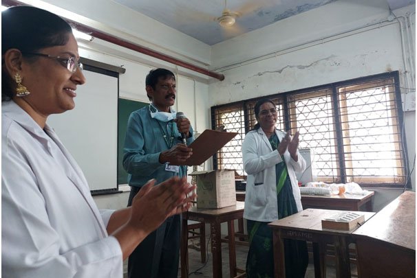
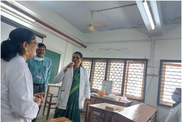
Judges: Prof. Dr. Thenmozhi, M.D., Vice Principal — Prof. Dr. P. Kannan, HOD of Cardiology — Prof. P. Sumathi, Professor in Surgery
Publications — Dept. of Anatomy, GMKMC, Salem‑30
| S.No | Name of Staff | Date of Publication | Topic | Journal |
|---|---|---|---|---|
| 1 | Dr. Kalai. Anbusudar, M.D, Associate Professor | 20.07.2016 (Accepted); December 2016 (Published) | Anatomical Study on Variations of Fissures of Lung | IJCAP (Indexed) |
| 2 | Dr. Kalai. Anbusudar, M.D, Associate Professor | 12.01.2016 (Accepted); 31.01.2016 (Published) | A Study of the Bifurcation of Sciatic Nerve & Its Clinical Importance | IJAR (Indexed) |
| 3 | Dr. Kalai. Anbusudar, M.D, Associate Professor | Ongoing Study (as part of ACME) | A Comparative Study Between Dissection Method and Video-Based Method of Teaching in Anatomy for I MBBS Students | — |
| 4 | Dr. Kalai. Anbusudar, M.D, Associate Professor | 25.10.2013 | Anatomical Study on Intraparotid Course of Facial Nerve and Its Terminal Branches | JEMDS (Indexed) |
| 5 | Dr. Kalai. Anbusudar, M.D, Associate Professor | 10.06.2013 | A Common Coeliaco-Mesenteric Trunk and Variations in Their Branching Pattern — A Case Report | JEMDS (Indexed) |
| 6 | Dr. Kalai. Anbusudar, M.D, Associate Professor | 10.06.2013 | A Very Rare Variation of the Superior Mesenteric Artery — A Case Report | JEMDS (Indexed) |
| 7 | Dr. Kalai. Anbusudar, M.D, Associate Professor | 14.11.2018 | A Study of the Origin of Superior Thyroid, Lingual and Facial Arteries | IJA (Indexed) |
| 8 | Dr. Kalai. Anbusudar, M.D, Associate Professor | 01.01.2020 | An Anatomical Study on Branching Pattern of Coronary Arteries — A Cadaveric Study | IJA (Indexed) |
| 9 | Dr. M.R. Manimekalai, M.D, Assistant Professor | March–April 2017 (Published) | A Study of Anatomical Variations in the Origin and Branching Pattern of the Superior Mesenteric Artery | RJPBCS (Indexed) |
| 10 | Dr. M.R. Manimekalai, M.D, Assistant Professor | 31.01.2017 (Published) | A Prospective Analysis of Tear Drop Angle in Intra-Articular Distal Radius Fractures Treated by Volar Buttress Plating | IJOTS (Indexed) |
| 11 | Dr. C. Sasikala, M.D, Tutor | Ongoing Study | An Anatomical Study on Branching Pattern of Axillary Artery — A Cadaveric Study | — |